Research
Research Focus
Translational Neuroscience

We are well established in translating models of disease from the research laboratory to the bedside of the patient. This important step levitates the model research towards a clinical setting, and therefore we use human intended techniques and devises. In translational research we always have focus on future treated patients.
The anatomy and physiology of the pig resamples that of a human. The Göttingen minipig keeps a relative low bodyweight, allowing both chronic and acute models.
By using our well-established large animal models to mimic human disease, we are able to test new devises, experimental treatment paradigms and imaging modalities.
Our approach on translational research is defined by both our own laboratory, and in collaborations with inhouse and external groups.
The portfolio of translational research stretches from preclinic GLP- (Good laboratory practice) and GCP- studies (Good clinical practice) to method and model development.
Our experimental facility is located inside the hospital, only steps away from the clinic. This ensures an easy access to collaborations, and a close contact to both patients and medical staff.
For us “bench to bedside”, is not just a saying. It is our reality.
Animal models of neurologic and psychiatric diseases
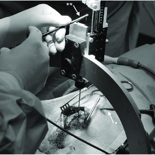
Large animal neuroscience studies the nervous system in the cat, dog, sheep, pig and primate, and offers several advantages compared with small animal neuroscience. The larger animal brain size enables the use of conventional clinical brain imagers and the direct use and testing of surgical procedures and equipment from the human clinic. The greater complexity of the large animal brain additionally enables a more direct translation to human brain function in health and disease and complement promising small animal basic studies by constituting an intermediate research system, bridging small animal CNS research to the human CNS. Large animal models may, accordingly, be of uttermost importance in the field of translational neuroscience.
Neuromodulation
The CENSE group has during the last 25 years used the Göttingen minipig to examine neuromodulatory treatment modalities such as stem cell transplantation and deep brain stimulation directed towards neurodegenerative diseases. The advantage of the Göttingen minipig is that it has a large gyrencephalic brain that can be examined at sufficient resolution using conventional clinical scanning modalities. The large brain, furthermore, enables the use of deep brain stimulation (DBS) electrodes and other neuromodulatory devices for human use, making the animal ideal for preclinical tests.
The primate, mice and rat models have resolved important aspects of the pathophysiology of neurodegenerative disease, but also have limitations. For example, all primate models are hampered by economical and ethical considerations leading to inclusion of few animals. Experimental rodent models on the other hand exclude, due to the small size of the rodent brain, use of conventional MR- and PET-imaging as well as use of the actual DBS devices intended for human use.
Neurosurgery
Our research in Neurosurgery focuses on the major neurosurgical diseases, on patient treatment, and developments in patient care and logistics.
Cancer in the brain
Cancer in the brain is major focus area and a multidisciplinary team specialises in treating all types of brain tumours. Treatment of cancer in the brain has been optimised by the department over the last five years with new techniques and patient logistics as well as database registries.
Spine surgery
Spine surgery is another focus area of the department where especially the cervical spine is a core competence of the department. The department treats degenerative diseases in the cervical spine as well as in the thoracic and lumbar spine. The department also takes part in the treatment of spine fractures and spinal cord injuries. The department performs treatment for acute spinal cord injury in collaboration with The Spinal Cord
Stroke
The Department of Neurosurgery is part of the Stroke Centre at Aarhus University Hospital. The department excels in the treatment of intracerebral bleedings both due to spontaneous hypertension or cerebral aneurysms. The department is responsible for intracranial/extracranial bypass treatment in patients with complex neurovascular conditions.
Neuromodulation
Neuromodulation treatments in patients with movement disorders such as Parkinson’s disease, essential tremor and dystonia was started in 1996 as the first department in Denmark and the treatment has remained leading and has led to innovation within electrode types and patient logistics.
The department houses the Center for Experimental Neuroscience (CENSE) which focuses on development of large animal models for neurological, neurosurgical, and psychiatric disorders. The animal models serve as a test platform for new treatments and technical developments within the neurological and neurosurgical specialities.
The Department of Neurosurgery has established an interdisciplinary research team to strengthen the neurosurgical area of specialisation through interdisciplinary research and development. Work is carried out to ensure and further develop the active research environment in collaboration with Danish Neuroscience Center.
The interdisciplinary research team works to make the neurosurgical research visible through publication in scientific journals and through presentation in printed and electronic media. We work to strengthen the collaboration between the university-based research units and other relevant research units, both nationally and internationally.
Neuromodulation
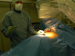
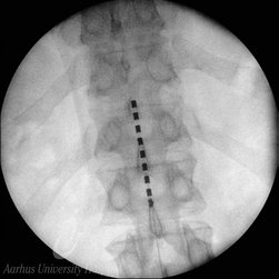
Modulation of the brain and nerves is one of our cornerstones. We have experience with stereotaxic precise targeting of both cortical areas, subcortical structures and single nerves. By modulation of the brain and nerves, it is possible to create new models of disease, test new treatment modalities in models, and treat medical conditions. We are currently working with intrathecal delivery, viral vector mediated overexpression of genes, lesion in substructures using electrical currency for ablation, stem cell therapy, deep brain stimulation, transcranial magnetic stimulation, radiation modulation, MR-guided focused ultrasound and spinal cord stimulation. Our research portfolio is based on both basic research, preclinical research and clinical trials. We are working with neurodegenerative diseases, movement disorders, chronic pain, cancer and psychiatric conditions. Neuromodulation is a fast-growing field, that constantly sprouts new methods and targets. CENSE-group is not only following the trends of research, but are in many fields the spearhead. We are adding on, redefining and refining our platform in order to find and solve neuromodulation tasks. Furthermore, using advanced electrophysiology, we are able to get biofeedback, that can be used to modulate neuromodulation in vivo, which is an important part of man-computer-interface.
Pain Research
We have worked clinically and scientifically with spinal cord stimulation (SCS) for chronic, neuropathic pain for more than 15 years. The last 5 years we have also worked with occipital nerve stimulation (ONS) for chronic cluster headache (Horton’s Headache), and with peripheral nerve stimulation (PNS) for painful mononeuropathies.
Our research has been focused on investigating predictors for treatment outcome, mechanisms of action, cost/benefit modeling, safety in pregnancy and outcome parameters. The Neurizon Neuromodulation Database, founded in 2012, forms the cornerstone of our research and registers all patients treated with neuromodulation for chronic pain in Western Denmark.
Spine Research
Spinal cord injury:
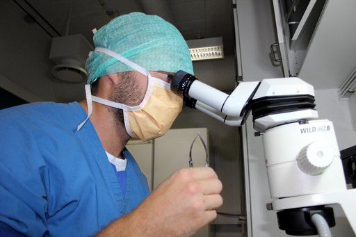
Our mission is to improve everyday functioning, quality of life, reduce incapacities and relieve handicaps for the individual who will suffer a traumatic spinal cord injury. We focus on strengthening treatment options within the earliest hours after onset of traumatic spinal cord injury. The avenues include the cascades of cellular, biochemical, inflammatory and immunological pathophysiology. In parallel we aim to address concomitant structural changes, microvascular changes, ischemia, hypoxia, axonal deterioration as well as the regenerative potential of the traumatized spinal cord. Essentially, we aim to reduce or even obliterate the primary and secondary pathophysiological processes in individuals suffering from a traumatic spinal cord injury.
Neuroanatomy & Neurobiology
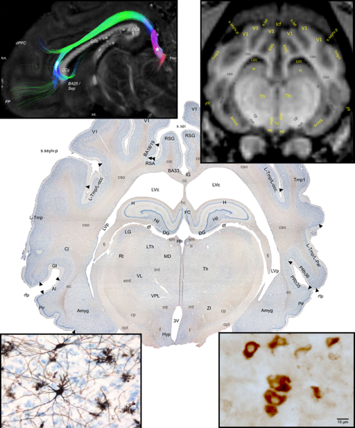
Preclinical studies and disease modelling are not possible without a thorough knowledge about brain anatomy and physiology. Therefore, an important part of CENSE group activity is studying the neuroanatomy and histology of the brain of our model animals, with a focus on Göttingen minipigs. Portfolio of the techniques used by us to describe the anatomy includes neuroimaging (MRI imaging, MRI-DWI tractography), reconstruction of the brain connectivity using stereotactically injected neuronal tracers (e.g. fluorogold or BDA) and various histochemical or immunohistochemical methods. Our experiments require good knowledge of the targeted structures, therefore we described in details various areas of the Göttingen minipig brain (like the subthalamic nucleus, substatia nigra, hypothalamus, septum, nucleus accumbens and so on), what, together with Nissl stained sections, allowed us to construct and publish first Gottingen minipig brain atlas, freely available online. The atlas includes also described MRI images and serve as a useful tool for researchers working with minipigs. The experiments performed by our group often require a post-mortem tissue analysis, therefore, we are constantly working on the development of the techniques for minipig brain removal and sectioning, histological staining and subsequent tissue analysis (using both quantitative (stereology and image analysis) and qualitative methods). Our group use those methods to describe nervous tissue response in various conditions, like diseases modelling, testing new devices (e.g. DBS electrodes), or new treatment paradigms (e.g. implanted stem cells).
Neurooncology
Neuro-oncology encompasses a wide spectrum of cancer diseases in the brain from primary brain cancers to metastatic disease. At the CENSE neuro-oncology division, our overall mission is to prolong and improve the lives of patients with cancer in the central nervous system. Our focus is on innovative projects with translational potential, and our research is aimed at the following three main areas of interest.
- Tumor treating fields therapy (TTFields): Tumor treating fields is a new and promising treatment of glioblastoma and other extra cranial cancers, which uses low intensity alternating fields to inhibit cancer growth. At CENSE Neuro-Oncology, we developed novel computational technique for estimation of the TTFields dose. The technique was developed in collaboration with the Danish Research Center for Magnetic Resonance and uses individualized MRI based computational models to provide and accurate representations of the tumor inhibiting field distribution throughout the brain and tumor. This can be used to improve clinical treatment planning, patient selection and prediction of treatment response. Our preclinical work has translated into two institution sponsored clinical trials testing novel dose-enhancing implementations for better patient outcome.
- Molecular biology and biobank: Since 2013, we have systematically collected tissue from numerous CNS tumors for the Danish Regional Cancer Biobank with the prospect of histopathological and molecular genetic research. Currently, our efforts are focused on proteome analysis from tears and tissue samples as novel tool in non-invasive monitoring in malignant brain tumors. This project is conducted in collaboration with the Danish Cancer Society and has the perspective of allowing early detection of brain cancer and recurrence.
- Database registries: Our clinical epidemiology research is currently work in progress focused on investigating the effects of novel diagnostic and treatment technologies used in our institution. Specifically, we are building a database with data from numerous diagnoses from benign to malignant CNS tumor in the period 2010 until 2035. The database will comprise a wide range of data, including clinical data, volumetric tumor measurements, 2HG spectroscopy measurements, and 18-FET-PET data, as well as data on the surgical techniques including intraoperative neuromonitoring and mapping, pre-surgical mapping (fMRI, MEG, nTMS) and fluorescence-guided methods, such as fluorescein or 5-ALA administration.
Techniques/Infrastructure
Stereotaxic Neurosurgery and Neuronavigation
Stereotaxic and neuronavigated neurosurgery is a minimally invasive form of surgical intervention which makes use of a 3D dimensional coordinate system to locate targets in the brain and spine.
Stereotactic and neuronavigated neurosurgery is comprised of:
1) A stereotactic planning system, including neuroanatomical atlas multimodality image matching tools, coordinates calculator, etc.
2) A stereotactic device or neuronavigation apparatus
3) A stereotactic / neuronavigation localization and placement procedure
All modern stereotactic and neuronavigation planning systems are computer based.
The stereotactic atlas is a series of cross sections of anatomical structure, depicted in reference to a two-coordinate frame. For example by a series of MR images. Based on this each brain structure is assigned a range of three coordinate numbers, which will be used for positioning the stereotactic device.
In most navigation systems the three dimensions are: latero-lateral (x), dorso-ventral (y) and rostro-caudal (z).
In order to perform MRI guided stereotaxic surgery in the minipig, a MR Localizer box and stereotaxic system has been developed by the CENSE group. The system combines the MR compatibility of its MR localizer box with a standard steel parallel rail stereotaxic frame and a high precision X,Y,Z micromanipulator. The animal is fixed in the frame by titanium screws penetrating into the zygoma bone in combination with a hard palate snout holder. The design preserves stereotaxic accuracy through the MRI and the stereotaxic procedures. The design allows easy neurosurgical access to the brain.
Anaesthesia and surgical procedures in large animal models
The size of the Göttingen minipig brain permits the use of full-scale neurosurgical equipment and advanced imaging modalities similar to those used in humans.
Anaesthesia: The pigs will first be lightly sedated with midazolam (20 mg, i.m.) for transportation to the CENSE experimental neurosurgical facility at Aarhus University Hospital. On arrival, the pigs are further sedated by a subcutaneous injection of a mixture of midazolam (0.5 mg/kg, Roche) and ketamine hydrochloride (10 mg/kg, Parke-Davis). A dose of buprenorfin (0.3 mg, Reckitt and Colman) is given via an indwelling catheter in an ear vein or IM. After induction of operative anesthesia, pigs will be intubated and maintained on assisted ventilation (according to weight) with an air mix with 2% Servoflurane. Circulatory homeostasis is maintained with continuous infusion of isotonic saline (20ml/h) and 5% glucose (20 ml/h). During the surgical procedure and the following MRI or PET procedures, physiological functions (heart rate, O2 saturation, expired air CO2, ECC) are monitored and recorded continuously. Body temperature is maintained in the normal minipig range (37.5-38.C) using a thermostatically controlled heating blanket.
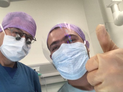
Neurosurgery: The minipigs are placed under general anesthesia as described above. Using routine sterile surgical technique, a sagittal skin incision in placed in the scalp. Using a high-speed drill, the calvarium is opened to the dura. The dura is opened using a neurosurgical micro-lancet and access to the brain is ready. Interventions can be injections of neural tracers, stem cells, viral vectors, neurotoxins or microbiological organisms, as well as, implantation of devices such as electrodes, micro chips or infusion devices or catheters. After the neurosurgical procedures the skin incisions are sutured by sterile technique. In order to prevent wound infection prophylactic intravenous injection of 750 mg cefuroxime is given at the start of the surgery and just before termination of the anesthesia. The pigs are then allowed to regain consciousness before being returned to the animal care facility. Post-operative pain therapy is treated administered with three times daily intramuscular injection of 5 mg buprenorphine for 3 days.
Neurostereology and neurohistology
Our research activity often requires a post-mortem analysis of the nervous system tissue. This may be basic research concerning Göttingen minipig anatomy, translational studies and disease modelling or verification of the new surgical techniques (like testing new brain-machine interfaces, verification of the precision of the stereotactic surgery etc.). Handling of the relatively large (in comparison with rodents) minipig brain is not simple. Therefore, CENSE group developed several techniques for animal perfusion, brain extraction, tissue slicing, freezing and processing using various histological techniques. Our neuro-histo-lab has extensive experience with processing of the nervous tissue using a variety of techniques, for frozen and paraffin-embedded samples. In many cases, the qualitative description of the histological sections is not sufficient; therefore, we are using also quantitative methods. Image analysis allows us to estimate several parameters describing the histological images, like area covered by the staining, morphology of the cells (using various shape descriptors) or changes in the dendritic spine density. Our group have also experience with stereology, used for unbiased estimation of the number of cells or fibers as well as volumes of various brain parts.
Deep Brain Stimulation (DBS)
Deep brain stimulation (DBS) is a neuromodulation technique based on surgical placement of electrodes in regions of interest in the brain. The electrodes are connected to a computer device allowing stimulation in different patterns and at different electric currents. By using electric stimulation in the brain, it is possible to bypass, enhance or “mute” certain electrical biological and neurochemical pathways. DBS is used to treat many different types of ailments, starting in the 1980´ies with movement disorders and later chronic pain, psychiatric and cognitive disorders. As DBS can modulate the brain, it is also possible to use for enhancement of different kinds of brain functions e.g. memory. By using devises that not only stimulate, but also record electrophysical activity, it is possible to modulate the stimulation. Some patients might need extra stimulation when they are doing exercise, and less when they are sleeping. We are working with DBS in clinical treatments, clinical experiments, translational models and devise testing.
Spinal Cord Stimulation (SCS)
See Pain Research above.
Behavioural analysis in large animal models
In order to verify the effect of treatment or examine neurological symptoms in translational models, we are using a variety of different test paradigms. Inspection and evaluation of a neurological symptom can be both easy and tricky. We have a neurological examination for pigs, but in order to quantify and correlate differences we use well defined methods. Activity, behavioral patterns and speed is being recorded and analyzed using a ceiling mounted camera. Differences in gait can be monitored and quantified using a pressure-sensitive gait mat system. In cases with unilateral differences in the brains gait function, rotational tests can be performed, either by monitoring spontaneous rotation, or by drug enhanced rotation e.g apomorphine or amphetamine. Animals are trained by special educated animal staff, in order to complete tasks, wear jackets with pockets for test equipment and walk on pressure sensitive gait mats.
Neuroimaging
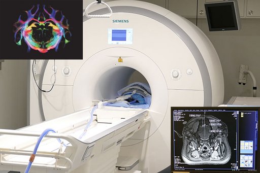
Cense group have extensive experience with the use of various imaging techniques in neuroscience-related research using large animals (pigs and minipigs) under anesthesia. The size of the pig/minipig brain allows us to use standard clinical imaging modalities. Routinely, we are using anatomical and stereotactic MRI in the planning of the surgeries and evaluation of their outcome (verification of the electrode placement, effects of the electrocoagulation etc.) as well as in the long term studies (like the evaluation of the effect of radiation on the nervous tissue). Designed by our group MRI compatible localizer box and stereotactic frame allows us to precise targeting of various minipig brain structure. We have also experience with ex-vivo, post mortem MRI and fiber reconstruction. Other imaging techniques used by us include computed tomography (CT), positron emission tomography (PET - used in the analysis of the brain activity or dopaminergic neurotransmission studies) or X-ray imaging (fluoroscopy). Our group have established close collaboration with the PET Centre and Center of Functionally Integrative Neuroscience (CFIN) MRI group, localized in the Aarhus University Hospital, allowing us to test various imaging paradigms.